हमें कॉल करें now
07971189475
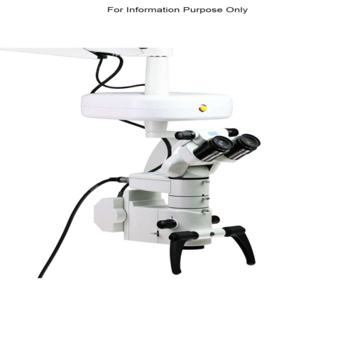

Specification
- स्पीड रेंज
- Variable Zoom & Focus
- सटीकता
- High Precision Optics
- विशेषताएँ
- LED Illumination, Adjustable Magnification, Modular System
- डिस्प्ले टाइप
- Binocular Viewing Head
- तापमान प्रतिरोध
- Ambient
- शेप
- Ergonomically Designed
- बिजली की आपूर्ति
- Integrated Electrical Power Unit
- कंट्रोल टाइप
- Motorized & Manual
- ग्लास टाइप
- High-Quality Optical Glass
- टाइप करें
- आयाम (एल* डब्ल्यू* एच)
- Approx. 600 x 600 x 1400 mm
- प्रॉडक्ट टाइप
- Laboratory Inverted Microscope
- उपकरण सामग्री
- Aluminum Alloy, High Impact Plastic
- पावर
- 90 VA
- वोल्टेज
- 100-240V AC, 50/60 Hz
- मटेरियल
- Metal & Optical Glass
- एप्लीकेशन
- Ophthalmic Surgery & Procedures
- Observation Tube
- Binocular, 45 inclined
- Working Distance
- 200 mm
- Illumination Type
- LED with adjustable intensity
- Light Source
- Cold light LED
- Camera Port
- Integrated for digital documentation
- Arm Extension
- Rotating and extendable arm
- Motorized Focus
- Vertical movement with joystick control
- Anti-Reflective Coating
- Yes
- Field of View
- Wide field, up to 25 mm
- Operating Modes
- Bright field, Optional Fluorescence
- Filter System
- Built-in UV and IR protection filters
- Magnification Range
- 6x, 10x, 16x, 25x, 40x, 63x
- Weight
- Approx. 90 kg
Trade Information
- Minimum Order Quantity
- 1 टुकड़ा
- मुख्य निर्यात बाजार
- , , , , , , ,
Tell us about your requirement

Price: Â
Quantity
Select Unit
- 50
- 100
- 200
- 250
- 500
- 1000+
Additional detail
मोबाइल number
Email

अधिक Products in अस्पताल उपकरण Category
इनोर्बविक्ट हेल्थकेयर इंडिया प्राइवेट लिमिटेड लिमिटेड
GST : 27AADCI6120M1ZH
GST : 27AADCI6120M1ZH
|
दुकान नंबर 311, तीसरी मंजिल, जिओन मॉल, हिंजवडी,पुणे छावनी - 411027, महाराष्ट्र, भारत
फ़ोन :07971189475
Mr. Rajesh Meshram (Director)
Mob: 08068820202, + 91 9850558881
Mob: 08068820202, + 91 9850558881
- अस्पताल उपकरण
- Hospital Equipment
- नैदानिक उपकरण
- Medical Equipment
- Hospital Equipment
- न्यूरोलॉजी मशीन
- अल्ट्रासाउंड मशीन
- दंत चिकित्सा उपकरण
- Medical Equipment
- आईसीयू और एनआईसीयू उपकरण
- डायलिसिस उपकरण
- न्यूरोलॉजी उपकरण
- Medical Equipment
- रेडियोलॉजी उपकरण
- क्लिनिकल डायग्नोस्टिक्स
- सी-आर्म मशीन
- बंध्याकरण उपकरण
- एंटीवायरल ड्रग्स
 जांच भेजें
 एसएमएस भेजें
 मुझे निःशुल्क कॉल करें
INORBVICT HEALTHCARE INDIA PVT. LTD.
सर्वाधिकार सुरक्षित.(उपयोग की शर्तें)
इन्फोकॉम नेटवर्क प्राइवेट लिमिटेड . द्वारा विकसित एवं प्रबंधित
इन्फोकॉम नेटवर्क प्राइवेट लिमिटेड . द्वारा विकसित एवं प्रबंधित

